MOLECULAR LANDSCAPES BY DAVID S. GOODSELL
Myelin
Acknowledgement: Illustration by David S. Goodsell, RCSB Protein Data Bank. doi: 10.2210/rcsb_pdb/goodsell-gallery-030
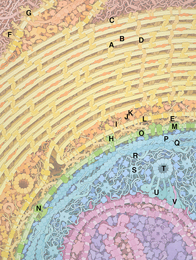
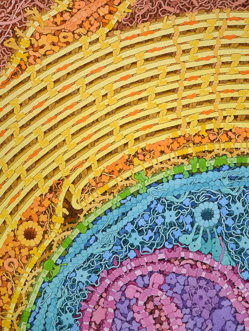
This painting shows a cross-section through the internode region of a myelinated axon in the central nervous system. The axon is shown at the bottom, with the membrane in green, the cytoplasm and cytoskeleton in blue, and a mitochondrion in purple and magenta. The oligodendrocyte wraps multiple times around the axon, shown with the membrane and membrane-bound proteins in yellow and the cytoplasm and cytoskeleton in orange. A small region of connective tissue is shown at the top in red.
Many structural and functional features are shown in the illustration, often requiring some artistic license:
- Proteolipid protein, myelin basic protein, and zig-zag stripes of claudin-11 glue the layers of myelin together.
- Myelin-associated glycoprotein on the inner surface of the oligodendrocyte reaches across and interacts with glycolipids on the axon membrane. More information on this protein is presented in a Molecule of the Month article.
- Myelin-oligodendrocyte glycoprotein on the outer surface of the oligodendrocyte makes a speculative interaction with proteoglycans.
- Voltage-gated sodium and potassium channels are shown in the axon membrane, although they are far more plentiful in the Nodes of Ranvier, rather than in the internode region shown here.
- A braided ring of actin supports the axon membrane, and successive rings are connected by spectrin.
- Neurofilaments and microtubules form a cytoskeleton inside the axon. Long intrinsically-disordered segments control the spacing between them.
- Mitochondria are transported by molecular motors like dynein along microtubules.
- A collection of soluble enzymes taken from a proteomic study fill the oligodendrocyte, including glycolytic enzymes, lactate dehydrogenase, fatty acid synthase and others. A similar collection is shown inside the axon.
- Monocarboxylic acid transporters may deliver lactic acid from the oligodendrocyte to the axon.
Myelin-specific proteins
A. PLP (proteolipid protein)
B. MBP (myelin basic protein)
C. MOG (myelin oligodendrocyte glycoprotein)
D. Claudin-11 (based on claudin-15)
E. MAG (myelin-associated glycoprotein)
Other Oligodendrocyte Proteins
F. Integrin
G. Dystroglycan complex
H. Signaling complex
I. Tyrosine-protein kinase Fyn (based on src)
J. Septin
K. Anillin
L. MCT1 (monocarboxylate transporter 1)
Axon
M. Voltage-gated sodium channel
N. Voltage-gated potassium channel
O. MCT2 (monocarboxylate transporter 2)
P. Actin
Q. Adducin
R. Spectrin
S. Neurofilament
T. Microtubule
U. Dynein-dynactin
V. Syntaphilin

Selected References
- Djannatian M, et al. (2019) Two adhesive systems cooperatively regulate axon ensheathment and myelin grown in the CNS. Nat Commun 10: 4794
- Vassilopoulos S, et al. (2019) Ultrastructure of the axonal periodic scaffold reveals a braid-like organization of actin rings. Nat Commun 10: 5803
- Stassart RM, Mobius W, Nave KA, Edgar JM (2018) The axon-myelin unit in development and degenerative disease. Front Neurosci 12: 467
- Yuan A, Rao MV, Veeranna, Nixon RA (2017) Neurofilaments and neurofilament proteins in health and disease. Cold Spring Harb Perspect Biol 9: a018309
- Kirkcaldie MTK, Collins JM (2016) The axon as a physical structure in health and acute trauma. J Chem Neuroanatomy 76: 9-18
- Nave KA, Werner HB (2014) Myelination of the nervous systems: mechanisms and functions. Annu Rev Cell Dev Biol 30: 503-533
- Scherer SS, Arroyo EJ (2014) Myelin: molecular architecture of CNS and PNS myelin sheath. Reference Module in Biomedical Research doi:10.1016/B978-0-12-801238-3.04676-6
- Xu K, Zhong G, Zhuang X (2013) Actin, spectrin, and associated proteins from a periodic cytoskeleton structure in axons. Science 339: 452-456
- Jahn O, Tenzer S, Werner HB (2009) Myelin proteomics: molecular anatomy of an insulating sheath. Mol Neurobiol 40: 55-72.
Learn more about other molecular landscapes:

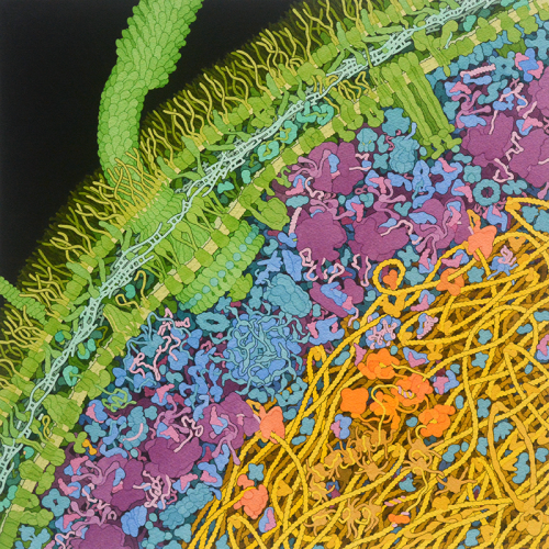
Escherichia coli Bacterium
This painting shows a cross-section through an Escherichia coli cell. The characteristic......

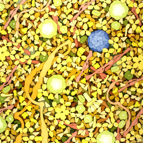
Biosites: Blood Plasma
This painting includes many Y-shaped antibodies, large circular low density lipoproteins......

